How do you do stain to evaluate sperm morphology?
For the correct visualization of a sperm sample it is important to know how to stain to evaluate sperm morphology. Approximately 15% of couples have infertility problems. Morphology is an important parameter for successful fertilization.
In assisted reproduction techniques (Kuster et al., 2004) the Kruger criteria (1986) and the WHO classification are the most important. According to the Kruger criteria, the head, neck and tail of the sperm must be normal and 70% of the head must be covered by the acrosome.
There are many stains available as Papanicolau, Hematoxiline, Eosine, Gimsa, Diff quik, etc. (Sanchez et al., 1994).
In this article we explain how you do the stain with Diff Quik.
How do you do stain to evaluate sperm morphology? Procedure :
1. Dispense Diff-Quik® Solutions into staining jars with lid or any other dip staining device.
2. Prepare the slides in the same way as for a Pappenheim technique (smear sample on a clean slide (put 10 µL for each smear and let air dry).
3. Dip slide 7 immersion or 1 minute in Fixative Solution (in Human, in other animal the time is different). Allow excess to drain each dip.
4. Dip slide 7 immersion or 1 minute in Stain Solution I (in Human, in other animal the time is different). Allow excess to drain after each dip.
5. Dip slide 7 immersion or 1 minutes in Stain Solution II (in Human, in other animal the time is different). Allow excess to drain after each dip.
6. Rinse slide with distilled water (gently)
7. Allow to air dry and proceed with differentiation. Look at the sample in the microscope. If you observe staining with low staining, repeat steps 4 and 5.
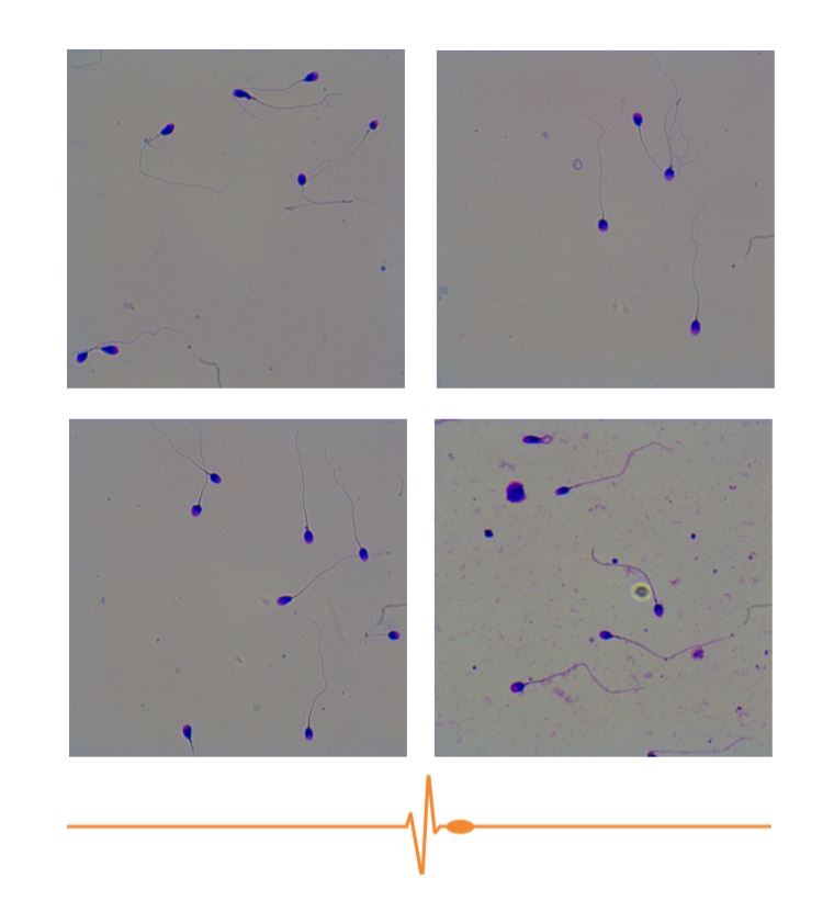
NOTE: If the sample is very concentrated, take into account that it is necessary to carry out an adequate dilution (that is similar to the images)